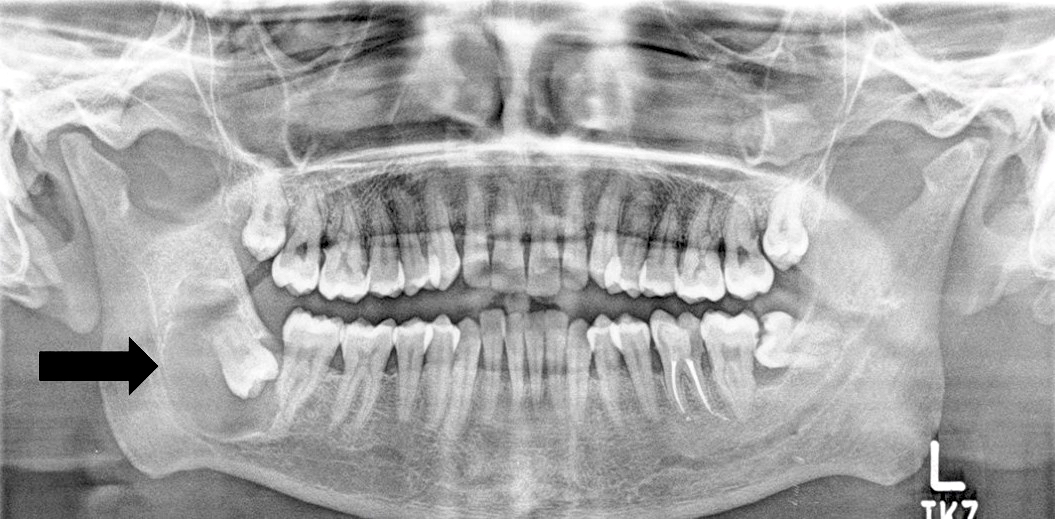
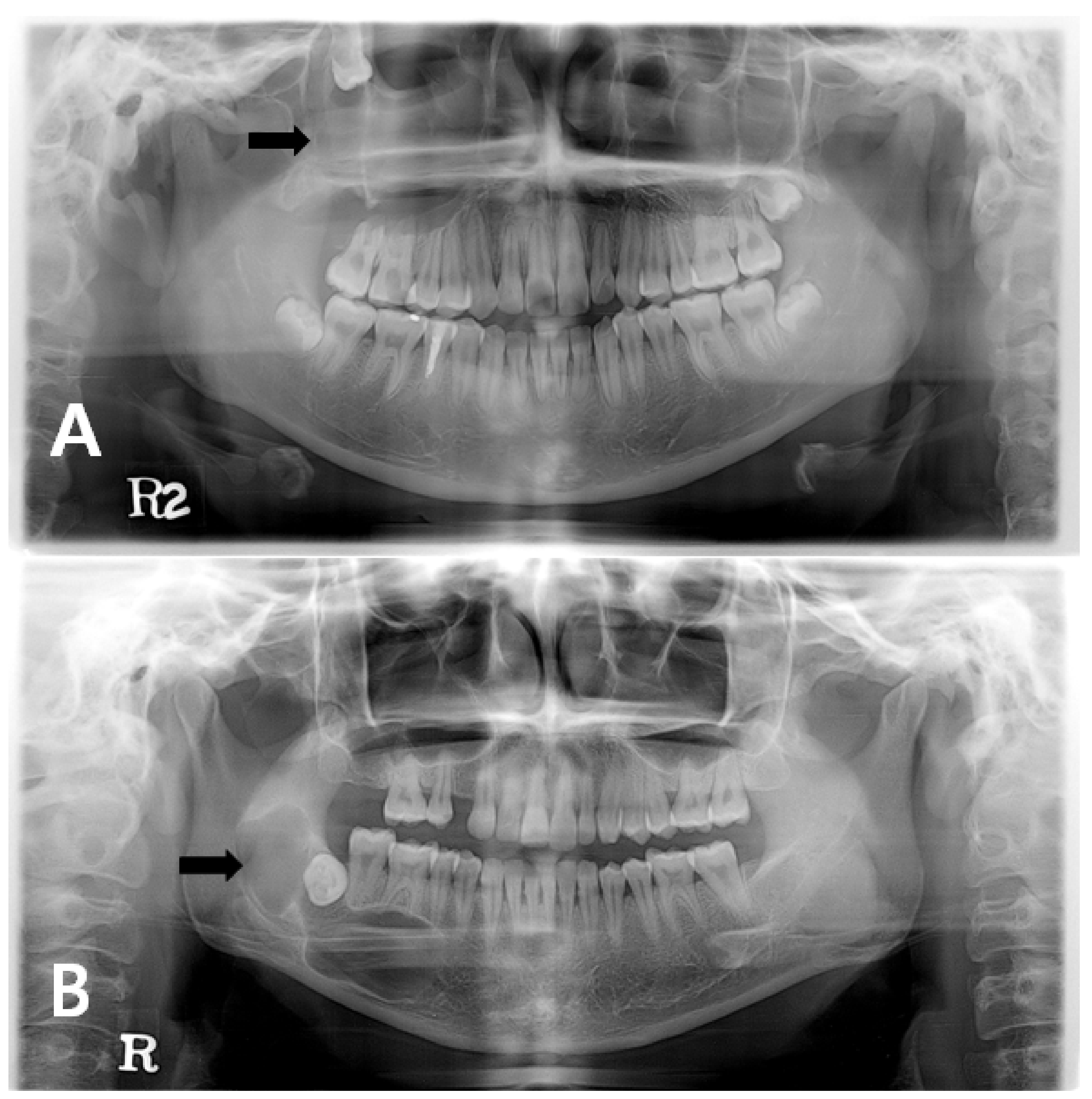
orthokeratinized odontogenic cyst
Orthokeratinized odontogenic cyst OOC is a relatively uncommon developmental cyst comprising about 10 of cases that had been previously coded as odontogenic keratocysts OKCs. To include as many reports as possible the emphasis was placed on recall rather than precision.
OOC exhibits distinctive clinical pathologic and behavioral features that varied substantially from KCOT and hence now it is considered as a separate entity.

. 2 the cartilaginous wall of odontogenic keratocyst was. Various studies have shown that OOC has typical characteristic clinicopathologic features when compared to other developmental odontogenic lesions such. The treatment of OOC is by enucleation and the prognosis following enucleation is excellent with a recurrence rate of less than 2.
In case 2 a 23-year-old woman presented a radiolucent lesion surrounded by. OOC had been previously coded as odontogenic keratocyst OKC and was termed as orthokeratinized variant of OKC. The terms used to search LILACS were keratocyst orthokeratinised odontogenic cyst and orthokeratinized odontogenic cyst.
We prefer to suggest our case unique collisions of two distinct simultaneously developing lesions orthokeratinized odontogenic cyst and cartilaginous heterotopia for the following reasons. In the WHOIARC classification of head and neck pathology this clinical entity had been known for years as the odontogenic. Orthokeratinized Odontogenic Cyst OOC is a developmental odontogenic cyst characterised by a lining of orthokeratinized stratified squamous epithelium 1.
Orthokeratinized odontogenic cyst OOC is a relatively rare odontogenic cyst characterized by the presence of orthokeratinized epithelial lining1 Here we reported an OOC that presented as a residual cyst at the right maxillary tuberosity in a 50-year-old female patient. However there had been controversy because the histological and clinical features of OOC and. Bearing in mind that both the MeSH for dentistry and radiology are generally inadequate and that free-text searching may not identify.
1 the epithelial lining did not demonstrate characteristics of odontogenic keratocyst. Orthokeratinized odontogenic cyst OOC is a rare developmental odontogenic cyst characterized by orthokeratinized stratified squamous epithelial lining. The orthokeratinized odontogenic cyst OOC is a developmental odontogenic cyst relatively rare arising from the cell rests of the dental lamina 1 2.
Recognition of OOC as a unique entity has long been due yet its inexplicable radiographic presentation resembling dentigerous cyst histological likeness to odontogenic keratocyst OKC and inconsistent cytokeratin expression profiles overlapping with both as well as with the. The orthokeratinized odontogenic cyst OOC is a rare developmental jaw cyst. The keratocystic odontogenic tumor which was previously referred to as odontogenic keratocyst must be differentiated from other odontogenic cysts because of its aggressive behavior.
Keratocystic odontogenic tumors can be seen at any age but are most common between 10 and 40 years of age in males and within the posterior mandible. Background Orthokeratinized odontogenic cyst OOC a newly designated entity of odontogenic cysts is an intraosseous jaw cyst that is entirely or predominantly lined by orthokeratinized squamous e. An odontogenic keratocyst is a rare and benign but locally aggressive developmental cystIt most often affects the posterior mandible and most commonly presents in the third decade of life.
Orthokeratinized odontogenic cyst OOC was first described by Schultz in 1927 and in 1945 Philipsen considered it to be a variant of Odontogenic keratocyst OKC. The treatment of OOC is by enucleation and the prognosis following enucleation is excellent with a recurrence rate of less than 2. OOCs were first described in 1927 by Schultz 2 as a variant of odontogenic keratocysts now known as keratocystic odontogenic tumours KCOTs 3.
Odontogenic keratocysts make up around 19 of jaw cysts. They were first identified by Wright in 1981 2 and were originally thought to be part of the spectrum of Odontogenic Keratocyst OKC 3. It was first described by Schultz in 1927 3 as an orthokeratinized variant of the formerly called odontogenic keratocyst today known as the keratocystic odontogenic tumour.
Orthokeratinized odontogenic cysts are a rare type of odontogenic cyst which are identified by an orthokeratinized stratified squamous epithelium. Orthokeratinized odontogenic cyst OOC is an odontogenic cyst was initially termed as the uncommon orthokeratinized type of odontogenic keratocyst by the World Health Organization. It usually occurs in mandible.
A benign developmental odontogenic cyst mostly unilocular with a fibrous tissue wall lined predominantly or entirely by orthokeratinized stratified squamous epithelium Essential features Cystic bone lesion in mandible or maxilla with compatible radiological features Cyst has orthokeratinized stratified squamous epithelial lining. Orthokeratinized odontogenic cyst OOC is a rare intraosseous cyst characterized by an orthokeratinized epithelial lining and minimal clinical aggressiveness 1. These cysts were originally classified as a subtype of odontogenic keratocysts.
Orthokeratinized Odontogenic Cyst OOC is a rare developmental odontogenic cyst which was considered in the past to be a variant of Odontogenic keratocyst OKC later renamed as keratocystic odontogenic tumor KCOT. However they have been redefined as a distinct entity. The histopathological examination revealed an orthokeratinized odontogenic cyst.
Orthokeratinized odontogenic cyst OOC is a developmental cyst of odontogenic origin and was initially defined as the uncommon orthokeratinized variant of odontogenic keratocyst OKC. 16 In 1981 Wright 2 reported 59 cases of what he then termed orthokeratinized variant of OKC which showed little clinical aggressiveness. Orthokeratinized Odontogenic Cyst OOC is a rare developmental odontogenic cyst which was considered in the past to be a variant of Odontogenic keratocyst OKC later renamed as keratocystic odontogenic tumor KCOT.

Pin By Neha On Oral Pathology Oral Pathology Pathology Oral

Pdf Orthokeratinized Odontogenic Cyst A Case Report A Milder Variant Of Okc Or An Independent Entity Semantic Scholar

The Photomicrograph Shows Cystic Lumen Lined By A Continuous Layer Of Download Scientific Diagram

Pathology Outlines Orthokeratinized Odontogenic Cyst

Pin By Neha On Oral Pathology Oral Pathology Dentistry Pathology

Pdf Orthokeratinized Odontogenic Cyst A Report Of Two Cases In The Mandible

Figure 2 Orthokeratinized Odontogenic Cyst A Report Of Three Clinical Cases

Jcm Free Full Text Changes In Cellular Regulatory Factors Before And After Decompression Of Odontogenic Keratocysts Html

Pdf Glandular Odontogenic Cyst Associated With Ameloblastoma Case Report And Review Of The Literature Semantic Scholar

A Case Report Of Synchronous Ameloblastoma And Odontogenic Keratocyst Of The Mandible Belknap 2022 Oral Surgery Wiley Online Library

Orthokeratinized Odontogenic Cyst

Pdf Orthokeratinized Odontogenic Cyst

Orthokeratinized Odontogenic Cyst Stratified Squamous Epithelial Download Scientific Diagram

Odontogenic Cysts Pocket Dentistry
Journal Of Dental Research Orthokeratinized Odontogenic Cyst An Unusual Histopathological Presentation
Orthokeratinized Odontogenic Cyst Critical Appraisal Of A Distinct Entity


Comments
Post a Comment